Coronal section of heart labeled – The coronal section of the heart, a cross-sectional plane dividing the heart into anterior and posterior portions, offers a unique perspective for understanding cardiac anatomy and function. This article explores the anatomical landmarks, clinical applications, advanced imaging techniques, and future directions of coronal heart imaging.
The coronal section provides a comprehensive view of the heart’s chambers, valves, and vessels, enabling clinicians to assess cardiac structure and pathology with greater precision.
Anatomy of the Heart’s Coronal Section

A coronal section of the heart is a vertical cut through the heart that divides it into right and left halves. It provides a cross-sectional view of the heart’s internal structures and allows for the visualization of the chambers, valves, and vessels.
Chambers
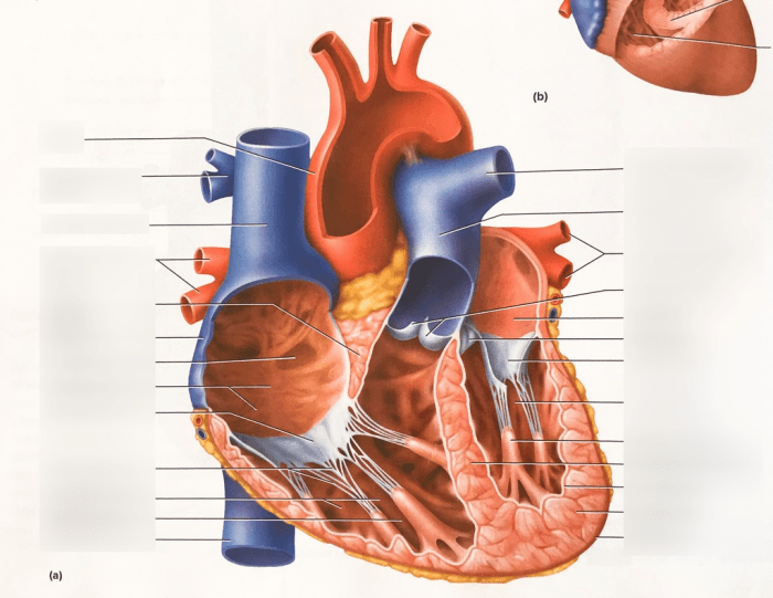
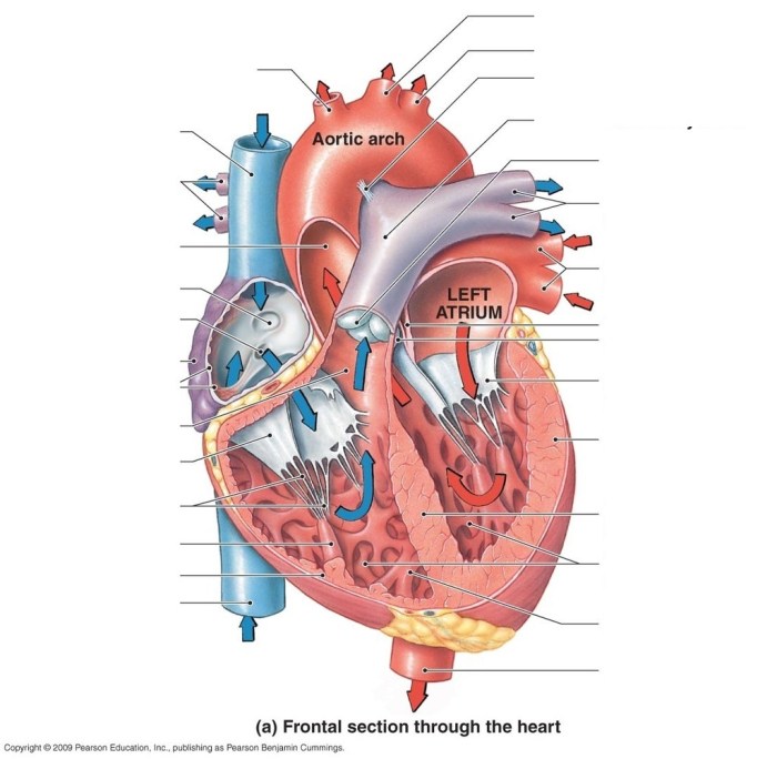
The coronal section reveals the four chambers of the heart:
- Right atrium:The right atrium is the receiving chamber for deoxygenated blood from the body. It is located in the upper right portion of the heart.
- Right ventricle:The right ventricle is the pumping chamber that propels deoxygenated blood to the lungs for oxygenation. It is located below the right atrium.
- Left atrium:The left atrium is the receiving chamber for oxygenated blood from the lungs. It is located in the upper left portion of the heart.
- Left ventricle:The left ventricle is the pumping chamber that propels oxygenated blood to the body. It is located below the left atrium.
Valves
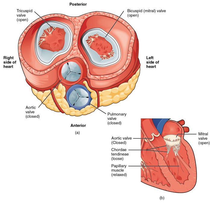
The coronal section also shows the four heart valves:
- Tricuspid valve:The tricuspid valve separates the right atrium from the right ventricle.
- Pulmonary valve:The pulmonary valve separates the right ventricle from the pulmonary artery, which carries blood to the lungs.
- Mitral valve:The mitral valve separates the left atrium from the left ventricle.
- Aortic valve:The aortic valve separates the left ventricle from the aorta, which carries blood to the body.
Vessels
The coronal section further reveals the major blood vessels associated with the heart:
- Superior vena cava:The superior vena cava is a large vein that carries deoxygenated blood from the upper body to the right atrium.
- Inferior vena cava:The inferior vena cava is a large vein that carries deoxygenated blood from the lower body to the right atrium.
- Pulmonary artery:The pulmonary artery is a large artery that carries deoxygenated blood from the right ventricle to the lungs.
- Aorta:The aorta is the largest artery in the body and carries oxygenated blood from the left ventricle to the body.
Clinical Applications of Coronal Heart Imaging

Coronal heart imaging offers a unique perspective of the heart’s anatomy and function, providing valuable insights for diagnosing and managing cardiac conditions.
The coronal section allows visualization of the heart’s chambers, valves, and coronary arteries from a different angle compared to traditional axial or sagittal views. This enables the identification of abnormalities that may be missed in other imaging planes.
Cardiac Function Assessment
- Coronal imaging is useful for evaluating cardiac function, including ejection fraction, wall motion abnormalities, and regional myocardial perfusion.
- It provides a comprehensive view of the heart’s motion and allows for the detection of subtle changes that may indicate underlying pathology.
Valvular Heart Disease
- Coronal imaging is particularly valuable in assessing valvular heart disease, as it allows for the visualization of the valves from multiple angles.
- It helps in diagnosing valve stenosis, regurgitation, and other valvular abnormalities, guiding treatment decisions.
Coronary Artery Disease
- Coronal imaging can be used to assess coronary artery disease by visualizing the coronary arteries and identifying stenoses or blockages.
- It provides information about the location and severity of the disease, aiding in the planning of interventions such as angioplasty or bypass surgery.
Advanced Imaging Techniques for Coronal Heart Analysis

Advanced imaging techniques play a pivotal role in obtaining detailed coronal images of the heart. Two prominent modalities in this regard are computed tomography (CT) and magnetic resonance imaging (MRI).
Computed Tomography (CT)
CT involves using X-rays and computer processing to generate cross-sectional images of the heart. It offers excellent spatial resolution, enabling visualization of anatomical structures with high precision.
Advantages:
- High spatial resolution for detailed anatomical assessment
- Rapid acquisition time, reducing motion artifacts
- Widely available and relatively cost-effective
Limitations:
- Exposure to ionizing radiation
- Lower soft tissue contrast compared to MRI
- Potential for motion artifacts in patients with arrhythmias or respiratory motion
Magnetic Resonance Imaging (MRI)
MRI utilizes magnetic fields and radio waves to generate images of the heart. It provides excellent soft tissue contrast, allowing for detailed visualization of cardiac structures and function.
Advantages:
- Excellent soft tissue contrast for distinguishing different cardiac tissues
- Non-invasive and does not involve ionizing radiation
- Ability to assess cardiac function and blood flow dynamics
Limitations:
- Longer acquisition times compared to CT
- Susceptibility to motion artifacts
- Higher cost and less widespread availability
Advanced imaging techniques have significantly improved the accuracy and efficiency of coronal heart analysis. They enable detailed assessment of cardiac anatomy, detection of abnormalities, and evaluation of cardiac function. These techniques have revolutionized the field of cardiology, providing valuable insights for diagnosis, treatment planning, and patient management.
Future Directions in Coronal Heart Imaging: Coronal Section Of Heart Labeled

Coronal heart imaging continues to advance rapidly, driven by technological innovations and the increasing availability of advanced imaging techniques. Emerging technologies, such as deep learning and artificial intelligence (AI), hold promise for further enhancing the diagnostic and prognostic capabilities of coronal heart imaging.
AI algorithms can be trained to analyze large datasets of coronal heart images, identifying patterns and relationships that may not be apparent to the human eye. This can improve the accuracy and efficiency of disease detection, risk stratification, and treatment planning.
Potential Applications of AI in Coronal Heart Imaging
- Automated detection and quantification of coronary artery disease
- Assessment of myocardial viability and function
- Prediction of cardiovascular events
- Personalization of treatment strategies
Areas for Further Research and Development, Coronal section of heart labeled
Despite the significant progress made in coronal heart imaging, there are still areas where further research and development are needed to enhance its utility.
- Standardization of imaging protocols:Establishing standardized imaging protocols will ensure consistency and comparability of images across different institutions and imaging systems.
- Development of new imaging biomarkers:Identifying and validating new imaging biomarkers will improve the diagnostic and prognostic accuracy of coronal heart imaging.
- Integration with other imaging modalities:Combining coronal heart imaging with other imaging modalities, such as echocardiography and nuclear medicine, can provide a more comprehensive assessment of cardiac structure and function.
- Clinical implementation and validation:Translating research findings into clinical practice requires rigorous validation and implementation studies to demonstrate the clinical utility and cost-effectiveness of new imaging techniques.
Key Questions Answered
What structures are visible in the coronal section of the heart?
The coronal section reveals the atria, ventricles, valves, and major blood vessels, including the aorta, pulmonary artery, and superior and inferior vena cava.
How is coronal heart imaging used clinically?
Coronal imaging is valuable for diagnosing and managing conditions such as valvular heart disease, congenital heart defects, and cardiomyopathies.
What advanced imaging techniques are used for coronal heart analysis?
Computed tomography (CT) and magnetic resonance imaging (MRI) are commonly used to obtain high-quality coronal images of the heart.